|
Chronic
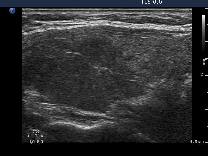
lymphocytic thyroiditis - Figure 2. Focal hypoechogenic areas.
Hypoechogenic areas within echonormal or minimally hypoechogenic background - this is most common sonographic presentation of Hashimoto's thyroiditis. The correct interpretation of this pattern is the essence of thyroid ultrasonography. The most important properties in the differentiation of thyroiditis and a nodule in pathological sense are the following: the basic echpattern, the sum, the size and the borders of the hypoechogenic lesions and the consideration of clinical and biochemical data. A clear distinction is possible in around 80% of such patterns. |
CLOSE |
|
Around 30% of the thyroid is hypoechogenic. These discrete lesions are not of regular geometric and have a characteristic puzzle-like, sharp, irregular borders. |
|
 |
 |
|
Aorund 50% of the thyroid is hypoechogenic. In the left, horizontal section hypoechogenic fields are in echonormal background, while in the left, longitudinal section, echonormal islets are in hypoechogenic background. The borders between the echonormal and the hypoechogenic areas are sharp and irregular. |
|
 |
 |
|
Hypoechogenic fields are much larger in this case. The irregular shape and the presence of smaller, moderately hypoechogenic areas are of help in avoiding overinterpretation these lesions as nodules. |
|
 |
 |